HIT News (School of Physics/ text) On July 13, Associate Professor Wang Jian of the School of Physics of our institute published the latest research results on the topic of Airy-Beam Tomographic Microscope in the latest issue of Optica, an authoritative international optical journal. He proposed a new non-scanning, high-resolution, three-dimensional microscopic imaging technology ATM based on Airy light field and successfully applied it to biological cell imaging. Optica is the flagship journal of the American Optical Society (OSA). Associate Prof. Wang Jian is the first author and co-author of the paper.
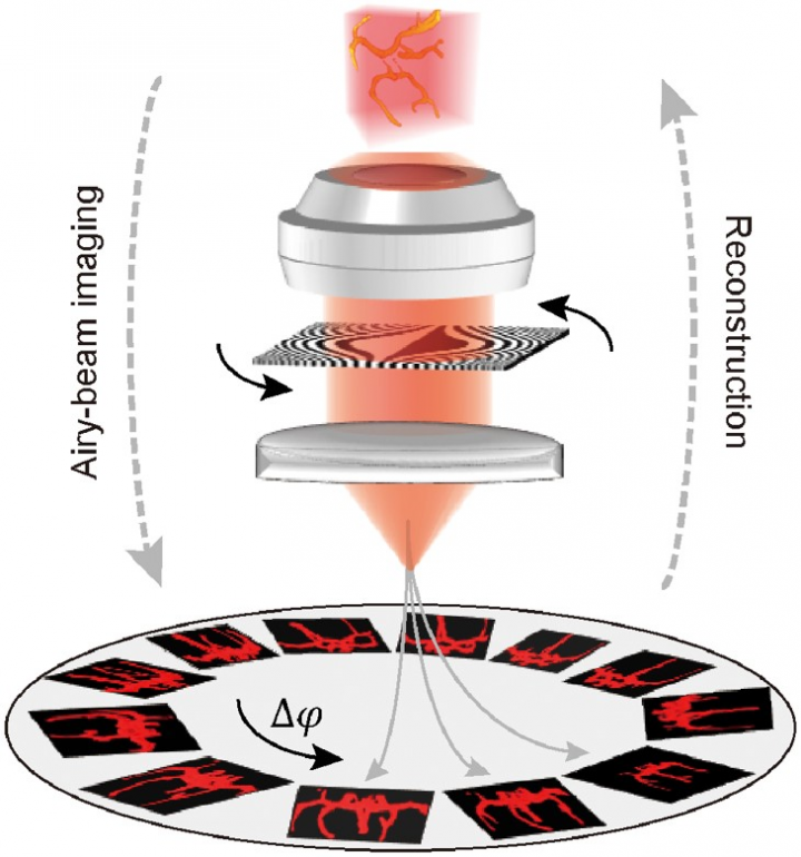
After frequency domain modulation, Gaussian distribution light field can generate Airy beam with non-diffraction, self-repair and self-acceleration characteristics. The non-diffraction characteristic of the beam is helpful to improve the resolution of optical imaging. The self-repairing characteristic can reduce the scattering influence of light beam passing through the medium and improve the imaging signal-to-noise ratio. The self-acceleration characteristic can realize the transverse self-bending propagation of the beam in free space. Applying the above characteristics comprehensively, the team proposed a unique three-dimensional microscopic imaging method ATM (see Fig. 1), which is based on two-dimensional projection image reconstruction. Only by changing the pattern on the modulator, high-resolution three-dimensional target images can be reconstructed without mechanical scanning.

Fig.1 Principle of ATM Imaging
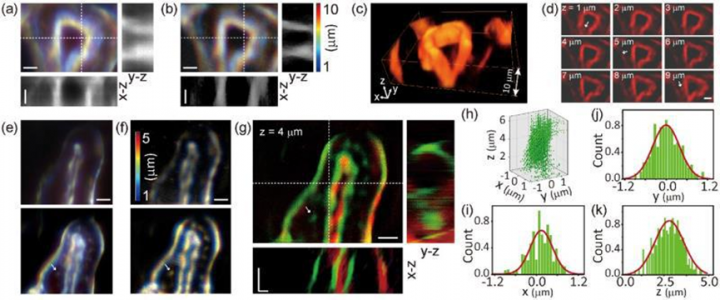
ATM imaging process includes many innovative technologies such as propagation control of Airy beam, PSF control and projection reconstruction algorithm. Chirp processing in frequency domain is used to increase the propagation distance in one side of focal plane and suppress the influence of beam sidelobe on imaging resolution. Particle imaging experiments show that the lateral resolution of the technology is 400-700nm and the depth resolution is 1-2 microns under 40 times objective lens. In this paper, the renal tubes and glomeruli in mouse renal cells were observed by this technology (see Fig. 2). Compared with the traditional Z-scan imaging technology, ATM technology boasts the advantages of high signal-to-noise ratio and deep field of view (above 10 microns) imaging without mechanical scanning. Combined with Airy beam three-dimensional reconstruction imaging algorithm, ATM lateral resolution is close to optical diffraction limit, and super resolution is realized in depth direction. This technology is expected to be applied in other 3D imaging technologies.

Fig. 2 (a): conventional Z-scan imaging of renal duct; (b): ATM imaging of renal ducts; (c) and (d): three-dimensional structure diagram and section diagram of renal duct by ATM image; (e):two-color Z-scan imaging of glomeruli (upper 568nm; lower 488nm); (f):two-color ATM imaging of glomeruli (upper 568nm; lower 488nm); (g): a two-color composite image; (h): an enlarged view of that tubular structure of figure (g); (i), (j) and (k): three dimension half-height width graphs of tubular structure.
Paper link:https://www.osapublishing.org/optica/abstract.cfm?uri=optica-7-7-790


